


Osteoarthritis of the knee joint is a specific disease that has a destructive effect on the cartilage of the knee joint.
When such a disease arises, doctors observe serious disturbances in the processes of blood circulation in the bone vessels, as a result of which deformation and destruction of cartilage tissue occurs, the person feels severe pain that constrains any movement of his legs and knees, and observes swelling and changes in the appearance of the knees.
To prevent the development of serious consequences of gonarthrosis (deformation of the limb and ankylosis - immobilization of the joint), when only joint replacement surgery can help the patient, the disease must be promptly identified and treated.Moreover, treat not independently with folk methods and advertised remedies, but under the supervision of qualified doctors.
What are the reasons?
The mechanisms of arthrosis in medicine are usually divided into:
- Primary- which occurs in old age from the natural aging of body tissues and against the background of some factors that provoke this disease, such as obesity (10% of cases), heredity, and increased stress throughout life.
- Secondary- it accounts for 30% of all cases of arthrosis of the knee joint; it usually manifests itself after an injury, a fracture of the tibia, ruptured ligaments, or damage to the meniscus.Moreover, with such arthrosis of the knee joint, the symptoms of the disease in most cases appear after 3-4 years, but after a serious injury it is possible even after 2-3 months.
In 50-60% of cases, the cause of arthrosis of the knee joint is a spasm of the muscles of the anterior surface of the thigh.
What happens to the knee joint with arthrosis?
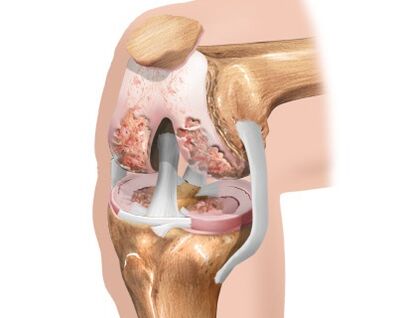
With excessive regular loads, genetic predisposition, metabolic disorders in the body, prolonged muscle spasms and injuries, the knee cartilage loses its smoothness and begins to thin out.The soft sliding of the articulated bones is replaced by strong friction, and the first degree of gonarthrosis develops, in which the cartilage loses its shock-absorbing properties.
The degradation process continues to progress, and poor shock absorption leads to flattening of bone surfaces with the formation of osteophytes in the form of bone growths.In this case, the disease already has a second degree and is accompanied by degeneration of the synovial membrane and joint capsule.Lack of pumping and movement atrophies the structure of the knee joint, the consistency of the knee fluid becomes more viscous, the process of nutrition of the cartilage is disrupted, which leads to an even greater deterioration of the patient’s condition.
Thinning cartilage leads to a reduction in the distance between articulated bones by up to 80%.While walking, due to abnormal friction and the absence of a shock absorber, destructive processes in the joint rapidly increase, this quickly leads to the development of the third degree of arthrosis with pronounced symptoms:
- Pain when moving, especially when going up or down stairs.
- Pain both during exercise and at rest, morning stiffness.
- The patient begins to limp, trying to spare the sore joint.
- Severe cases of the disease require the use of crutches or a cane.
The third degree of arthrosis of the knee joint is characterized by an almost complete absence of cartilage tissue, which leads to a decrease in joint mobility to a minimum.Therefore, no magical methods, super medicines or ointments can restore worn-out cartilage tissue, and given the degree of bone deformation, normal functioning of the joint is impossible.In this case, only surgery can help.
Stages of arthrosis
As arthrosis progresses, the following stages or degrees are distinguished:
- The initial stage, or stage 1, is often called compensated, since there are no pronounced clinical signs of pathology yet.The patient may feel discomfort in the knees after physical activity, which quickly disappears after rest;the joints are still fully functional.
- With degree 2 arthrosis, subcompensated, the symptoms of the disease intensify.A pronounced pain syndrome is formed, which, however, is relieved by anesthetic ointments and gels used topically.Motor activity is impaired and joint instability appears.In the vast majority of cases, patients turn to doctors at this stage.
- 3rd degree, decompensated, can also be called deforming.The knee joint is twisted, unstable, immobile, and completely non-functional.The pain syndrome is constant and requires serious medical intervention.To unload the joint and move, a person needs a cane.
Symptoms and first signs
Depending on the severity of symptoms, the development of the disease is divided into 3 stages.The symptoms of early development of gonarthrosis vary greatly and are not specific.
At the first stage of pathology, the following signs are possible:
- knee pain when squatting or walking up stairs;
- pain in the joint after prolonged exercise or cooling;
- the knee may hurt at the end of the day or in the morning;
- stiffness and soreness in the morning goes away after everyday work.
Subsequently, symptoms characteristic of stages 2 and 3 appear (in increasing order):
- the pain becomes acute and prolonged;
- swelling in the joint area;
- fluid may appear in the joint;
- between the articular surfaces, pinching of particles of cartilage, meniscus, synovial villi is possible, as a result of which sharp immobility occurs (it can also pass abruptly);
- it is difficult to step on your foot;
- joint immobility develops.
Consequences
If left untreated, the following complications of knee arthrosis may develop:
- Joint deformity.As a matter of fact, joint deformation is more likely not a complication, but the last stage of the disease.
- Infection in a joint.Infection usually occurs due to microtrauma.They can be considered small cracks in the cartilage tissue.Pathogenic microorganisms are introduced into the joint through the flow of blood or lymph from other areas.This often occurs after infectious diseases.
- Dislocations and fractures.These complications are explained by dysfunction of the knee joint.With arthrosis, there is no uniform distribution of load from the femur to the bones of the lower leg.The ligaments that normally strengthen the joint also weaken.Because of this, at a certain moment (even during normal walking) the bones of the lower leg may be subject to excessive stress, which will lead to a fracture or dislocation.To avoid this, people with severe structural and functional impairments in the knee joint should move with the help of a crutch or cane.
- Ankylosis.Ankylosis is the fusion of two bones where a joint once existed.This complication is perhaps the most severe, as the joint simply disappears.The tibia and femur gradually fuse, and the tibia is fixed in one position.Movement, of course, is impossible.
Diagnostics
To diagnose gonarthrosis use:
- blood tests (general and biochemical);
- radiography;
- arthroscopy;
- Ultrasound;
- MRI.
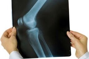
The most important diagnostic method for confirming the diagnosis of gonarthrosis is radiography.Although it is not possible to examine the initial stages of the disease and the state of the cartilage tissue on x-rays, nevertheless, bone changes of stages 2 and 3 are clearly visible.This:
- narrow joint space;
- marginal spines located along the contour of the bones, the edges of the patella - osteophytes;
- changes in the periosteum;
- change in the height of one of the condyles and others.
However, changes can be examined in more detail using arthroscopy.
Ultrasound and MRI help detect changes in the soft tissues of the knee joint during early arthrosis.These methods also provide a good indication of the condition of cartilage tissue, synovium and fluid.
How to treat arthrosis?
Treatment for arthrosis of the knee joint is long and sometimes painful.The duration is due to the fact that once the disease has manifested itself, it will constantly remind itself as long as the person lives.
Thus, in order to properly treat gonarthrosis, a fairly strong financial foundation and discipline are required.An important role is played by how seriously the patient takes treatment, since often, in order to reduce the frequency of exacerbations of the disease, the patient is forced to change his daily activity, favorite profession, engage in joint development, quit smoking and drinking alcoholic beverages.
There are three main stages in the treatment of deforming arthrosis of the knee joint.
The first stage of treatment includes:
- communicating to patients the essence of their disease, risk factors and secondary prevention measures;
- daily gymnastics with stretching elements;
- contrast shower;
- swimming in the pool 2 – 3 times a week;
- weight loss.
The second stage of treatment includes:
- external fixation of the joint using calipers, bandages, elastic bandages and orthoses;
- the use of ointments and creams based on non-steroidal anti-inflammatory drugs to relieve inflammation and pain;
- the use of drugs from the group of chondroprotectors to reduce the rate of cartilage destruction.
The third stage of treatment includes:
- course use of NSAIDs orally;
- intra-articular injections with hormonal anti-inflammatory drugs;
- additional use of drugs with a pronounced analgesic effect;
- surgical replacement of a diseased joint with an implant.
Along with drug treatment, modern medicine uses methods such as:
- Kinesitherapy.In this case, treatment of arthrosis of the knee joint is carried out using special exercises.The load is selected in accordance with the degree of development of the disease and the patient’s physical fitness individually.
- Ozone therapy.This is an effect on a sore knee joint using ozone.With this method of physiotherapeutic treatment, the substance can be administered by injection or used externally.
- dietary supplement.Biologically active supplements are a worthy alternative to other medications.
- Homeopathy.It involves taking medications in small doses.The course of treatment lasts only a few weeks, but during this time, provided that the drugs are properly selected, blood circulation and normal nutrition of cartilage tissue cells are completely restored.
- Exercisesallow you to improve blood circulation in the knee joint, restore the elasticity of the ligaments, improve the nutrition of cartilage tissue cells, and promote its restoration.
Drug treatment
All medications used in the treatment of gonarthrosis can be divided into those that help eliminate the main symptoms of the disease, and those that restore the function of the knee joint and prevent the progression of the pathology.The first type of drugs includes non-steroidal anti-inflammatory drugs and hormonal drugs.The second group includes chondroprotectors and hyaluronic acid.
Nonsteroidal anti-inflammatory drugs
Drug treatment of arthrosis begins with the elimination of pain.They are the ones who cause the greatest moral and physical suffering to patients and lead to loss of ability to work.Non-steroidal anti-inflammatory drugs (NSAIDs) have worked well among painkillers.
The drugs can be used in two ways - topically or orally (in tablets).Local treatments (preferred) often include patches, ointments, or gels.The analgesic effect, as a rule, occurs on days 3-4, and its maximum develops by days 7-10.
The course of treatment with nonsteroidal drugs should be limited to 10-14 days.NSAIDs have a limited range of use and are used with great caution in the treatment of elderly patients.
Hormonal drugs
In cases where NSAID treatment is not enough and the disease continues to progress, the doctor may prescribe hormonal injections.They belong to the “heavy artillery” means and help to quickly eliminate pain, relieve inflammation and swelling of surrounding tissues.
Due to the large number of side effects, hormonal drugs are prescribed in short courses, only during the acute period of the disease, when inflammatory fluid accumulates in the joint cavity.The drug is administered into the joint space no more than once every 10 days.
Chondroprotectors
To prevent further destruction of the cartilaginous surfaces of the joint, chondroprotectors are prescribed.
These drugs also help reduce pain and reduce signs of inflammation in the surrounding soft tissue.In addition, chondroprotectors slow down the destruction of articular tissues and stabilize the disease due to their shock-absorbing and lubricating functions.Chondroprotectors improve the nutrition of cartilage, normalize the composition and properties of intra-articular fluid, and protect pain receptors from excessive irritation.
Hyaluronic acid
Hyaluronic acid is called a fluid prosthesis of intra-articular fluid.In its properties, it is similar to natural synovial (intra-articular) fluid, which nourishes cartilage tissue and cushions the knee joint during movements.
Hyaluronic acid preparations are injected into the joint, thereby creating a thin protective film that prevents the cartilage surfaces from rubbing against each other.Injections are carried out only after the acute phase of the disease has passed.
Exercise therapy and exercises for arthrosis
Physical exercise helps restore joint function and strengthen it.They should be performed slowly and carefully so as not to cause sprains and not to exceed the permissible load.
The following exercises are considered the most effective:
- Slowly raise your straight legs one at a time while lying on your stomach.This exercise uses the thigh and calf muscles.The load should not be allowed to be transferred to the back.You shouldn't raise your legs too high either.The main thing is to tense your muscles at the top point;
- This exercise is similar to the first, only now you should lift your leg with the knee bent.At the top point, the thigh muscles should be further tensed.The exercise must be performed the same number of times on each leg;
- Lying on the floor (on your stomach), raise your straight legs up, and then spread them apart and bring them together.This exercise requires strong, trained abdominal muscles, so it is not suitable for all patients.In addition, performing it may increase blood pressure.For patients suffering from hypertension or other diseases of the cardiovascular system, it is better to avoid it;
- To perform the next exercise, you need to lie on your side, bend your leg lying on the floor at the knee, and slowly lift the other and hold it at the top point.It is important that the angle when performing on each leg is the same;
- You will need a chair for this exercise.Sitting on it, the legs are straightened one by one, raised up and held for the maximum possible time in a straightened position;
- It is useful to rise on your tiptoes while holding the back of a chair or bed.As with other exercises, you need to linger at the top point for a few seconds and additionally strain your leg muscles;
- Smooth movement from toe to heel allows you to activate blood circulation in the lower extremities.Such movements should be performed alternately: while one leg rests on the toe, the other on the heel and vice versa.All movements must be smooth;
- Leaning on the back of a chair or bed, stand on your heels for a minute, raising your toes up.If you cannot stand in this position for the specified time, you should start with less, gradually increasing it;
- A massage that is performed in a standing position.The legs should be rubbed with vigorous movements directed from the knee up to the thigh.It is necessary to complete the massage by stroking the skin.
The doctor will conduct several sessions in the office and show basic techniques so that the patient can massage the problematic knee on his own.Competent actions improve the condition of the damaged area.
Massage and self-massage
Perform self-massage with your palm, the edge of your palm, your fingertips, or your fist.Gently act on the sore knee, maintain forceful pressure on the affected area: strong patting, blows, active kneading of tissues often worsen the condition.
Basic techniques:
- the first stage is light stroking clockwise, the second stage is circular movements counterclockwise;
- Press one palm to the sore knee, lightly tap it with the fingertips of the other hand;
- maintain the initial position of the palm, tapping not with your fingers, but with the edge of your palm;
- gently stroke the knee in a circle, gradually increase the pressure (but in moderation);
- During therapy, massage the affected area twice a day for 10–15 minutes.For preventive purposes, it is enough to do a light massage once every 7 days.
Before the procedure, apply an ointment or gel with an anti-inflammatory effect to the sore knee.
Physiotherapeutic treatment
All methods of physiotherapy can be divided into several groups:
- Reducing pain;
- Reducing inflammation;
- Restoring nutrition to the joint and accelerating the restoration of joint function.
The doctor can determine what type of treatment the patient needs based on the existing symptoms and concomitant diseases.
Surgery for arthrosis
Surgical treatment refers to radical methods that partially or completely restore the functioning of the knee joint.Surgical treatment methods differ from each other in the degree of intervention in the affected joint.
- Arthroscopyrefers to the most gentle methods of surgical treatment.This technique is less traumatic and can be used as a therapeutic measure even in the early stages of arthrosis.Its main goal is to extend the life of the damaged joint.Arthroscopy is performed using an endoscope - a flexible probe with a camera at the end.Small punctures are made in the knee joint, through which an endoscope and auxiliary instruments are inserted.During the operation, damaged areas of tissue that cause pain are removed.The operation is most suitable for young people and, if necessary, can be repeated several times.
- Endoprostheticsrefers to the most radical surgical techniques.In this case, complete restoration of joint function occurs by replacing the entire knee joint or part of it with an implant.This method is the best alternative to the previously existing technique - arthrodesis (complete immobilization of the damaged joint).Currently, endoprosthesis replacement provides up to 90% positive results and significantly improves the quality of life of patients.
- OsteotomyIt is used when significant deformations have developed in the joint and the function of the joint is noticeably affected.Osteotomy involves creating an artificial bone fracture in a pre-planned location.Subsequently, the parts of the bone are aligned in the correct, physiological position and allowed to grow together.Sometimes during the operation, artificial fixators of bone fragments can be used, which contribute to a more stable position of the bone.
Folk remedies
Traditional medicine will also help you.The use of various lotions to relieve swelling and pain has long been practiced by many patients.Here are some useful recipes:
- Burdock leaves.Take 5 burdock leaves and hold them over boiling water to steam them.Lubricate the sore knee with vegetable oil and apply steamed leaves.Wrap your leg in cling film and a warm scarf.Leave the lotion for a couple of hours.
- Cabbage leaf lotion.It is made from fresh cabbage leaves soaked in May honey and applied to the leg.The therapeutic effect is strengthened by insulation made of woolen fabric or plastic film.This product is suitable for people of all age groups.
- Eggshells, kefir.Grind the shells of 2 eggs and mix with 1 spoon of kefir.Apply the mixture to your knee, wrap it with cloth and cling film, and lie down under a blanket.Leave it like this for a couple of hours, then rinse off the composition with water.This treatment can be used daily.
- Horseradish compress.According to this folk recipe, to enhance the medicinal properties of horseradish, it is recommended to first crush and boil it.Then it must be applied to the affected area.
- Turpentine– an excellent warming agent for sore joints.The knee is rubbed with turpentine before going to bed and tied with a woolen scarf.Already after the first procedure, the patient experiences significant relief.The course of treatment is selected individually for each person.
Diet
The therapeutic diet involves avoiding or minimally consuming canned, smoked and fried foods (to suppress appetite).In order to restore damaged cartilage, it is necessary to provide the body with complex carbohydrates (porridges, wholemeal products).Juices (carrot, beetroot, apple) should also be included in the diet.They will remove toxins from the body and reduce the impact of inflammatory processes.
The diet should include fish and aspic, which act as a kind of chondroprotector and help create new cartilage.It is worth remembering that you cannot prescribe a diet for yourself - only a nutritionist (diet specialist) can choose the best option.
Sample menu:
- Breakfast: oatmeal with water without butter or sugar, fruit juice, boiled egg;
- Second breakfast: a glass of low-fat natural yogurt;
- Lunch: steamed meat or fish, stewed vegetables, tea without sugar;
- Afternoon snack: cottage cheese casserole with nuts, a glass of fruit juice;
- Dinner: vegetable salad, apple, tea without sugar;
- Second dinner: a glass of low-fat kefir.
Prevention
Prevent joint diseases:
- dose the load on your legs during active sports;
- create a diet that includes foods and dishes containing calcium, magnesium, gelatin;
- if the work involves lifting or moving heavy objects, follow safety rules and do not carry loads exceeding a certain limit;
- Do self-massage regularly to prevent arthrosis, especially with constant stress on your legs;
- eat right, limit “harmful” foods;
- body weight control (excess weight means additional stress on the joints);
- Get regular examinations and treat acute and chronic diseases.
Forecast
Provided arthrosis of the knee joint is diagnosed in the early stages, the cause of the pathological process is eliminated and adequate treatment is provided, the prognosis is favorable.The therapy provided makes it possible to achieve long-term remission, but treatment is usually lifelong.
In the absence of the necessary treatment, as well as when the patient fails to comply with the doctor’s instructions, arthrosis of the knee joint becomes a cause of disability.